From the journals: JBC
A new mode of mammalian iron uptake. Self-made molecules that scatter Pseudomonas. Antibodies that ameliorate Alzheimer's. Read about recent papers in the Journal of Biological Chemistry on these and other topics.
A new way to absorb dietary iron
Iron, a mineral found in every cell of the human body, combines with proteins to form the hemoglobin required to transport oxygen. The body also needs iron for physical growth, neurological development and critical biochemical functions, making it an essential nutrient. However, iron in excess is toxic, and the body has no physiological means to excrete that excess. Instead, levels are regulated by iron absorption.
Dietary iron mainly exists in two forms: heme and nonheme. Plant-based and fortified foods are a source of nonheme iron, whereas meat, seafood and poultry supply both heme and nonheme iron. The National Heart, Lung and Blood Institute estimates that 20% of women, 50% of pregnant women and 3% of men have an iron deficiency, which is the most common cause of anemia, a decrease in red blood cells. This deficiency can be treated with iron supplements. However, absorption of available supplements is inefficient, and it often takes several months to reestablish normal iron levels.
Researchers recently identified nicotianamine-iron chelate, or NA-Fe2+, as a new form of bioavailable iron present in mice, chickens and plant-based foods. However, researchers know little about the mechanisms responsible for NA-Fe2+ absorption from its dietary sources. In a recent article in the Journal of Biological Chemistry, Yoshiko Murata and a team of Japanese researchers identified the proton-coupled amino acid transporter, or PAT1, as the principal channel through which NA-Fe2+ is absorbed. Using cultured cells modified to no longer contain the PAT1 protein, electrophysiological analyses of PAT1 overexpression in aquatic frog oocytes and experiments using mice, the authors demonstrated the uptake of NA-Fe2+ into the epithelium of the proximal jejunum and iron appearance in spleen, liver and kidney.
These findings confirm that NA-Fe2+ is a bioavailable source of iron, uncover the possible mechanism by which NA-Fe2+ is absorbed in the mammalian intestine, and may drive development of improved iron supplements.

A self-made molecule scatters Pseudomonas
The Gram-negative bacterium Pseudomonas aeruginosa causes hospital infections, particularly in patients with compromised host defense mechanisms, posing a significant health and economic burden. To become virulent, P. aeruginosa must attach to a surface such as epithelial tissue; it then forms a biofilm, whereby the pathogen secretes proteins that aid its spread. Researchers aim to identify agents that can disperse these bacteria and prevent them from propagating after they have attached.
Robert Scheffler and colleagues at Princeton University developed a quantitative single-cell surface-dispersal assay and demonstrated that P. aeruginosagenerates several factors that can stimulate its own dispersal. Using bioactivity-guided fractionation, mass spectrometry and nuclear magnetic resonance, the authors showed that one such factor, 2-methyl-4-hydroxyquinoline, inhibits the activity of type IV pili, or THP, the thin appendages that decorate P. aeruginosa, and leads to dispersal of the pathogen.
These findings, published in a paper in the Journal of Biological Chemistry, identify THP inhibition as a promising strategy against infection caused by P. aeruginosa.
Sorting out sinking phagocytosis
Phagocytosis, the receptor-mediated ingestion of cellular particles, is the major function of immune cells such as macrophages and neutrophils. Yet more than 100 years after Élie Metschnikoff — the scientist credited with discovering phagocytes — developed his theory of phagocytosis, scientists still know little about the processes that govern this cellular ingestion process. Researchers have described two structurally distinct modes of phagocytosis: phagocytic cup formation, whereby the membrane extends outward and engulfs the particle, and sinking phagocytosis, or inward engulfment of particles. A number of receptors that activate downstream signaling pathways initiate phagocytosis. One such receptor, CR3, is thought to drive sinking phagocytosis. However, evidence of this receptor's involvement is sparse.
In a recent study published in the Journal of Biological Chemistry, Stefan Walbaum and colleagues at the University of Münster in Germany isolated resident macrophages from normal unaltered mice and mice genetically altered to lack CR3 receptors to investigate how macrophages engage and internalize particles. Using RNA-sequence analysis, genetic experiments and real-time 3D imaging, the researchers showed that CR3 receptors indeed regulate a mode of sinking phagocytosis and also are involved in the formation and closing of Fc receptors for immunoglobulin G, or fragment crystallizable gamma receptor–controlled phagocytic cups that extend outward to engulf particles.
Ameliorating Alzheimer's with antibodies
The hallmark aggregates of the amyloid-beta, or Aβ, peptide that form in the brains of Alzheimer's disease patients display varying conformations. These different structures could have different pathophysiological consequences; for example, large, high–molecular-weight Aβ aggregates accelerate the accumulation of toxic Aβ fibrils.
In the past, researchers isolated two single-chain variable domain antibody fragments, or scFvs, called C6T and A4, which selectively bind to different conformations of Aβ aggregates and represent a promising therapeutic agent to selectively target Aβ variants.
Recent studies led by Ping He at Arizona State University use those scFvs to demonstrate that specific Aβ aggregate variants and their location influence the therapeutic response to scFvs in mice. The scFvs, which contained a peptide tag to facilitate transport across the blood-brain barrier, were administered to normal or Alzheimer's mice, and their effects were studied seven months later. C6T restored neuronal integrity in the Alzheimer's mice to nondiseased levels, promoted growth of new neurons and increased survival rates. Treatment with A4, on the other hand, decreased Aβ deposits but did not significantly decrease neuroinflammation or promote neuronal integrity, neurogenesis or survival.
These findings, reported in the Journal of Biological Chemistry, suggest that the specific Aβ conformation targeted in therapeutic applications may affect treatment efficacy — including attenuation of neuroinflammation and promotion of neuronal integrity —and that the location of the targeted Aβ variants also may be a critical factor.
The duality of coagulation factor V
Factor V, or FV, is a glycoprotein that has a dual role as both an anticoagulation and procoagulation cofactor. Central to its opposing functions is the proteolytic removal of its central B-domain, and researchers seek to understand the mechanisms surrounding this functional switch.
In a paper published in the Journal of Biological Chemistry, Teodolinda Petrilloand colleagues at the Children's Hospital of Pennsylvania describe their study of how bond cleavage in the B-domain influences interactions between FV and FV-short, a physiologically relevant isoform with a shortened B-domain, and tissue factor pathway inhibitor alpha, or TFPIα. Using clotting assays, chromatography, fluorescent anisotropy and biochemical analyses, the authors demonstrated that key forms of FV, including FV-short, are physiologic ligands for TFPIα. The authors speculate that strategies that target the interaction of TFPIα with these different forms of FV may provide a new way to strengthen or dampen the coagulation system.
Enjoy reading ASBMB Today?
Become a member to receive the print edition four times a year and the digital edition monthly.
Learn moreGet the latest from ASBMB Today
Enter your email address, and we’ll send you a weekly email with recent articles, interviews and more.
Latest in Science
Science highlights or most popular articles

E-cigarettes drive irreversible lung damage via free radicals
E-cigarettes are often thought to be safer because they lack many of the carcinogens found in tobacco cigarettes. However, scientists recently found that exposure to e-cigarette vapor can cause severe, irreversible lung damage.
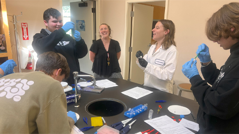
Using DNA barcodes to capture local biodiversity
Undergraduate at the University of California, Santa Barbara, leads citizen science initiative to engage the public in DNA barcoding to catalog local biodiversity, fostering community involvement in science.
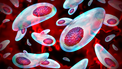
Targeting Toxoplasma parasites and their protein accomplices
Researchers identify that a Toxoplasma gondii enzyme drives parasite's survival. Read more about this recent study from the Journal of Lipid Research.
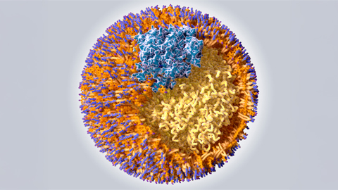
Scavenger protein receptor aids the transport of lipoproteins
Scientists elucidated how two major splice variants of scavenger receptors affect cellular localization in endothelial cells. Read more about this recent study from the Journal of Lipid Research.
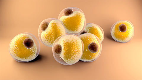
Fat cells are a culprit in osteoporosis
Scientists reveal that lipid transfer from bone marrow adipocytes to osteoblasts impairs bone formation by downregulating osteogenic proteins and inducing ferroptosis. Read more about this recent study from the Journal of Lipid Research.
Unraveling oncogenesis: What makes cancer tick?
Learn about the ASBMB 2025 symposium on oncogenic hubs: chromatin regulatory and transcriptional complexes in cancer.

