From the journals: JBC
Huntington protein interactions affect aggregation. Intrinsically disordered protein forms a scaffold. From unknown protein to curbing cancer growth. Read about papers on these topics recently published in the Journal of Biological Chemistry.
Huntington protein interactions affect aggregation
The neurodegenerative disorder Huntington’s disease arises from mutations in the exon1 region of the huntingtin protein, or Httex1. Aggregated Httex1 arise via fibril formation of Httex1 monomers and colocalizes with other aggregation-prone proteins like Tar-DNA binding protein, or TDP43. These aggregates can impede major cellular functions in the brain. In addition, TDP43 expression is associated with other neurodegenerative disorders, including amyotrophic lateral sclerosis. TDP43 aggregates by transitioning from liquid condensates to solid fibrils. Scientists want to understand the mechanisms behind Httex1 and TDP43 interaction and aggregation.
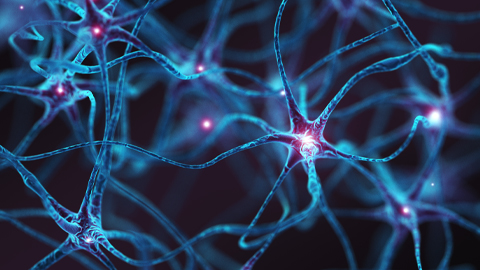
Gincy George and colleagues at the University of Southern California used biophysical, biochemical and cell biological techniques to determine that Httex1 and TDP43 directly interact. They published in the Journal of Biological Chemistry that the interaction is conformation-dependent, occurring between Httex1 fibrils and liquid condensates of the TDP43 low-complexity C-terminal fragment, or TDP43-LCD. This interaction expedites the liquid–solid transition and aggregation of TDP43-LCD, but it slows down Httex1 aggregation. Based on electron paramagnetic resonance spectroscopy data, the authors indicated that TDP43-LCD ties up Httex1 fibrils that can act as seeds to fuel further conversion of Httex1 monomers to aggregated fibrils.
Future studies will determine how the Httex1–TDP43-LCD interaction speeds up formation of solid TDP43-LCD and whether TDP43-LCD interacts similarly with other proteins or nucleic acids. Finally, the authors speculated that these findings could open new pathways to develop compounds that inhibit Httex1 aggregation and similarly mutated proteins to treat neurodegenerative disorders.
Intrinsically disordered protein forms a scaffold
Carboxysomes are bacterial microcompartments that contain proteins that drive CO2 fixation to sugars. These proteins include the enzymes ribulose-1,5-bisphosphate carboxylase/oxygenase, or Rubisco, and carbonic anhydrase, which both perform key reactions for carbon fixation; shell proteins; and a scaffolding protein. The scaffolding protein CsoS2 contains intrinsically disordered regions. Scientists found that intrinsically disordered proteins play important roles in signaling, regulation and macromolecular assembly. Therefore, understanding carboxysome assembly will help scientists engineer biological systems for industrial applications such as fuel and chemical production.
Julia B. Turnšek and colleagues at the University of California, Berkeley studied CsoS2 from the proteobacterium Halothiobacillus neapolitanus and published their findings on its contribution to carboxysome architecture in the Journal of Biological Chemistry. The authors found that conserved residues in the middle region of CsoS2 are essential for interacting with shell proteins.
In addition, the researchers determined that the CsoS2–shell protein interaction drives phase separation behavior. Using this finding, they artificially reconstituted spherical condensates containing Rubisco, CsoS2 and shell proteins. These condensates will be a useful biochemical tool for studying carboxysome assembly. Scientists still need to uncover how carboxysomes assemble into nano compartments and how they could be leveraged for industrial applications.
From unknown protein to curbing cancer growth
The WW domain-containing protein 1, or WWP1, is an E3 ubiquitin ligase that regulates the expression of proteins associated with cancer progression, such as members of the p53 superfamily. Increased WWP1 expression correlates with poor cancer prognosis, so scientists want to understand what factors regulate WWP1 levels. Using a yeast two-hybrid screen, Tiphaine Perron and colleagues from Sorbonne Université and other French institutes identified cysteine and tyrosine-rich protein 1, or CYYR1, as a WWP1 binding partner and regulator, giving function to this previously undefined protein in their recent publication in the Journal of Biological Chemistry.
Using coimmunoprecipitation experiments, the authors showed that PPxY motifs in CYYR1’s cytoplasmic tail are important for WWP1 binding. They also determined that CYYR1 induces WWP1 autoubiquitination where WWP1’s catalytic domain adds a ubiquitin chain to itself. This process leads to WWP1 degradation via the lysosome. Furthermore, the researchers found that CYYR1 limits breast cancer cell growth via its PPxY motif. Additional studies will characterize the CYYR1–WWP1 interaction in more detail and dig deeper into the possible role of CYYR1 in breast cancer tumor suppression.
Enjoy reading ASBMB Today?
Become a member to receive the print edition four times a year and the digital edition monthly.
Learn moreGet the latest from ASBMB Today
Enter your email address, and we’ll send you a weekly email with recent articles, interviews and more.
Latest in Science
Science highlights or most popular articles
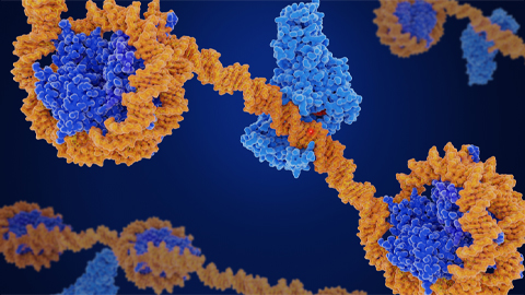
CRISPR epigenome editor offers potential gene therapies
Scientists from the University of California, Berkeley, created a system to modify the methylation patterns in neurons. They presented their findings at ASBMB 2025.
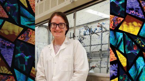
Finding a symphony among complex molecules
MOSAIC scholar Stanna Dorn uses total synthesis to recreate rare bacterial natural products with potential therapeutic applications.

E-cigarettes drive irreversible lung damage via free radicals
E-cigarettes are often thought to be safer because they lack many of the carcinogens found in tobacco cigarettes. However, scientists recently found that exposure to e-cigarette vapor can cause severe, irreversible lung damage.
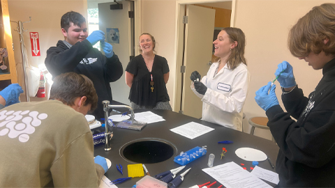
Using DNA barcodes to capture local biodiversity
Undergraduate at the University of California, Santa Barbara, leads citizen science initiative to engage the public in DNA barcoding to catalog local biodiversity, fostering community involvement in science.
Targeting Toxoplasma parasites and their protein accomplices
Researchers identify that a Toxoplasma gondii enzyme drives parasite's survival. Read more about this recent study from the Journal of Lipid Research.
Scavenger protein receptor aids the transport of lipoproteins
Scientists elucidated how two major splice variants of scavenger receptors affect cellular localization in endothelial cells. Read more about this recent study from the Journal of Lipid Research.

