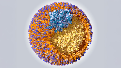
Structure of the key protein for an HCV vaccine


The hepatitis C virus, or HCV, causes a chronic liver infection that can lead to permanent liver scarring and, in dire cases, cancer. It affects around 71 million people worldwide and causes approximately 400,000 deaths each year. While treatments are available for HCV-related infections, they are expensive, hard to access and do not protect against reinfection. A vaccine that can help prevent HCV infection is a major unmet medical and public health need.
One major reason there hasn’t been an HCV vaccine yet is that scientists have yet to identify the proper antigen, or the part of the virus would trigger a protective immune response in the body.
Decades of research have pinpointed HCV E1E2, the only protein on the surface of the virus, as the most promising vaccine candidate. However, developing an HCV vaccine based on that protein is limited by uncertainty around what it looks like. Knowing the structure of the protein is necessary to figure out how the immune system responds to the virus.
So how do researchers capture the structure of single protein on a shape-shifting virus?
We are researchers who specialize in microscopy and vaccine design. With new technology, we were able to visualize the molecular details of this elusive protein, unlocking key insights into how this virus works and offering a potential blueprint for a future vaccine.
This is how we did it.
Challenges of capturing a shape-shifting virus
One reason it has been so difficult to capture the structure of the HCV E1E2 protein is that it is both flexible and fragile. It changes its shape so often and is so easily broken that it’s challenging to purify.
As an analogy, imagine a bowl of spaghetti drenched in tomato sauce. Now imagine trying to take a picture of each individual piece of spaghetti in the same position over time while the bowl is shaking. Hard to do, right? That’s what it was like to image the full E1E2 protein.
There were also technological barriers. Until recently, available imaging techniques were limited in their ability to view microscopic proteins. X-ray crystallography, for instance, is unable to capture molecules that frequently change and shape-shift, like HCV. Moreover, other options, such as nuclear magnetic resonance spectroscopy, required cutting large parts of the protein or chemically manipulating it in a way that would transform its physiological state and potentially alter its function.
So to examine the structure of E1E2, we needed a way to extract and purify, stabilize and trap the entire shape-shifting protein into one configuration.
How to take a picture of protein
Cryo-EM, or cryo-electron microscopy, is a type of imaging technique that views specimens at cryogenic temperatures, in this case the boiling point of nitrogen: minus 320.8 degrees Fahrenheit (minus 196 Celsius). With temperatures that cold, ice freezes so quickly that it doesn’t have time to crystallize. That creates a beautiful glasslike frame around the protein of interest, allowing an unhindered view of every structural detail. Cryo-EM also requires very little protein to work, reducing the amount of material we would need to purify.
Winner of the 2017 Nobel Prize in chemistry and Nature magazine’s 2015 “Method of the Year” award, cryo-EM is superb for imaging biological macromolecules in their native, or natural, state in the aqueous environment of human blood. Cryo-EM was also pivotal for characterizing the structure of the COVID-19 virus and its variants.
So how do you take a picture of a protein?
First, we embedded the genetic code to make E1E2 in human cells in a petri dish so we would have sufficient amounts of protein to study. After purifying the protein, we plunged it into liquid ethane followed by liquid nitrogen. Liquid ethane is used to freeze the protein because it has a higher boiling point than liquid nitrogen. This means it is able to capture more heat before turning to a gas, allowing the protein to freeze much more quickly than it would in liquid nitrogen and avoid structural damage.
Once the protein was vitrified, or in a glasslike ice state, we were able not just to see its overall structure, but also to capture multiple individual configurations of the protein that it takes when it shape-shifts, including its less stable forms.
At this point, our protein was ready for its close-up. We employed a microscope that uses a beam of focused, high energy electrons and a very fancy camera that detects how the elections bounce off the protein’s surface. This created a 2D image that we then mathematically transformed into a 3D model. And that was how we got the coveted “close-up” of HCV’s surface protein.
Our next step was then to assess the location of each amino acid, or building block of the protein, in 3D space. Because every amino acid has a unique shape, we used a computer program that could identify each one in our 3D map. This allowed us to manually reconstruct a high-resolution model of the protein, one building block at a time.
A new tool to design an HCV vaccine
Our 3D map and model of the HCV E1E2 protein supports previous research describing its structure while providing new insights into features that will help pave the way for a long-sought vaccine design against this virus.
For example, our structure reveals that the interface between the two main parts of the protein is stabilized by sugars and hydrophobic patches, or areas that push out water molecules. This creates sticky binding hubs along the protein and keeps it from falling apart – a potential site for protective antibodies and new drugs to target.
Researchers now have the tools to design antiviral drugs and vaccines against HCV infection.
This article is republished from The Conversation under a Creative Commons license. Read the original article.
![]()
Enjoy reading ASBMB Today?
Become a member to receive the print edition four times a year and the digital edition monthly.
Learn moreGet the latest from ASBMB Today
Enter your email address, and we’ll send you a weekly email with recent articles, interviews and more.
Latest in Science
Science highlights or most popular articles

E-cigarettes drive irreversible lung damage via free radicals
E-cigarettes are often thought to be safer because they lack many of the carcinogens found in tobacco cigarettes. However, scientists recently found that exposure to e-cigarette vapor can cause severe, irreversible lung damage.
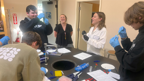
Using DNA barcodes to capture local biodiversity
Undergraduate at the University of California, Santa Barbara, leads citizen science initiative to engage the public in DNA barcoding to catalog local biodiversity, fostering community involvement in science.
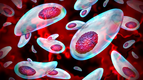
Targeting Toxoplasma parasites and their protein accomplices
Researchers identify that a Toxoplasma gondii enzyme drives parasite's survival. Read more about this recent study from the Journal of Lipid Research.
Scavenger protein receptor aids the transport of lipoproteins
Scientists elucidated how two major splice variants of scavenger receptors affect cellular localization in endothelial cells. Read more about this recent study from the Journal of Lipid Research.
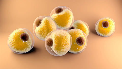
Fat cells are a culprit in osteoporosis
Scientists reveal that lipid transfer from bone marrow adipocytes to osteoblasts impairs bone formation by downregulating osteogenic proteins and inducing ferroptosis. Read more about this recent study from the Journal of Lipid Research.

Unraveling oncogenesis: What makes cancer tick?
Learn about the ASBMB 2025 symposium on oncogenic hubs: chromatin regulatory and transcriptional complexes in cancer.