From the journals: MCP
The long-term effects of limited oxygen in utero. How Alzheimer’s disease changes global proteome turnover. Spatial proteomics offer HIV-1 mechanism insights. Read about papers on these topics recently published in the journal Molecular & Cellular Proteomics.
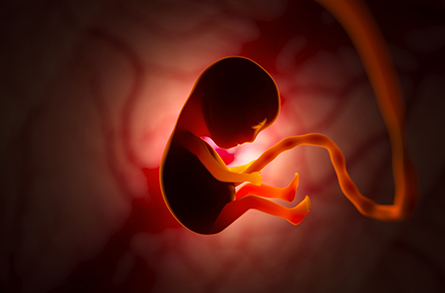
Limited oxygen in utero has long-term effects
Hypoxia is the state of insufficient oxygen in tissue, and chronic hypoxia refers to long-lasting low oxygen content in the blood. Hypoxia during pregnancy affects more than 2% of the world population, especially people living at high altitudes. It disrupts development of the embryo, which increases the risk of multiple chronic diseases, including diabetes and cardiovascular and chronic kidney disease, in adulthood.

In a recent paper in the journal Molecular & Cellular Proteomics, Stefan Rudloff at the University of Bern, Andrea Bileck at the University of Vienna and a team of researchers describe the effects of chronic hypoxia on mouse fetal kidneys. By comparing the proteomes of kidneys subjected to hypoxia and kidneys under normal oxygen conditions, they determined that hypoxia causes significant deregulation of proteins related to kidney development, metabolic adaptation and premature aging.
Nephrons are the structural and functional unit of the kidney. Under hypoxic conditions, fewer nephrons form, increasing the risk of renal dysfunction and failure in adulthood. Furthermore, chronic hypoxia causes neutrophils to accumulate at sites of nephron formation, causing the tissue damage and DNA oxidation observed in hypoxic fetal kidneys.
In response to insufficient oxygen, cells try to adjust metabolic pathways by shifting from oxygen-dependent oxidative phosphorylation to oxygen-independent glycolysis to extract energy from sugars. The researchers observed elevated levels of all 10 enzymes involved in glycolysis in hypoxic fetal kidneys. They found that the protein Klotho, which functions as both an intracellular anti-inflammatory and a systemic anti-aging factor, already was downregulated in fetal kidneys.
The authors concluded that the steps fetal cells take to survive hypoxia, instead of growing normally, can predispose tissues to faster aging in adulthood. Researchers can use the characteristic biomarkers of this accelerated aging to detect and target human diseases caused by chronic hypoxia.
Does Alzheimer’s affect global proteome turnover?
The rate of protein turnover in cells depends on the rate of protein synthesis and degradation. Depending on the tissue, proteins can have different turnover rates. For example, proteins in mammalian brains have a higher rate (minutes to days) than skeletal muscle proteins (typically weeks). Researchers believe that imbalances in protein turnover are a primary factor in common neurodegenerative diseases such as Alzheimer’s. Due to such imbalances, some neuronal proteins misfold and form aggregates that cannot be restored to their native form. Scientists do not yet know if neurodegenerative diseases affect proteome turnover beyond the brain.
In a recent article in the journal Molecular & Cellular Proteomics, Byron Andrews at the Medical Research Council Laboratory of Molecular Biology in the U.K. and a team of researchers describe their study of global proteome turnover at different stages of Alzheimer’s disease and amyotrophic lateral sclerosis, or ALS, in three transgenic mice models. Healthy mice were used as the control for the experiment.
The researchers’ results showed an increase in global average protein turnover at a later stage of disease in all three mouse models. This suggests that protein repair mechanisms are increasingly active as the disease progresses. The change in turnover was limited to the proteins of the forebrain, the region affected by Alzheimer’s disease. Turnover in serum and liver proteins was unaffected. In contrast to the disease models, global proteome turnover in healthy mice showed a steady decrease with aging. The authors attributed this to the slowdown of protein degradation through autophagy with aging.
Spatial proteomics offer HIV-1 mechanism insights
Cells are composed of organelles, compartments that perform specific tasks. Organelles require specific proteins to function, and a protein’s location within a cell provides valuable information about its function. This protein localization is a strictly regulated process.
To remain undetected by the cellular immune system and continue multiplying, viruses disrupt protein localization, leading to protein inactivation and cell dysfunction. Spatial proteomics provides information about protein localization in organelles and thus is a promising tool to understand the mechanism of disease progression at a subcellular level. Usual spatial proteomic analysis depends on reference organelle proteome; therefore, it cannot be used if proteins are translocated to different organelles. Researchers have not yet established which method is best to study cell disruptions caused by viral infection.
In a recent article published in the journal Molecular & Cellular Proteomics, Aaron L. Oom at the University of California, San Diego, and a team of researchers describe using machine-learning models to analyze protein localization in various organelles in human T lymphocyte cells containing an inducible HIV-1 genome. When these cells multiplied in the presence of an inducer (doxycycline in this case), the viral genome was expressed as well, and the HIV infection disrupted the subcellular localization of proteins.
The researchers analyzed spatial proteomics data of the host cell using three machine-learning models. Performance of these models depends on the organelle, meaning that for a particular organelle, one model performs better than the others. This result suggests that researchers should choose an appropriate machine-learning model to analyze spatial proteomics data of a specific organelle for the study of the HIV-1 infection mechanism. This work serves as a reference for the study of viral infections as well as additional mutants of the HIV-1 genome.
Enjoy reading ASBMB Today?
Become a member to receive the print edition four times a year and the digital edition monthly.
Learn moreGet the latest from ASBMB Today
Enter your email address, and we’ll send you a weekly email with recent articles, interviews and more.
Latest in Science
Science highlights or most popular articles

From humble beginnings to unlocking lysosomal secrets
Monther Abu–Remaileh will receive the ASBMB’s 2026 Walter A. Shaw Young Investigator Award in Lipid Research at the ASBMB Annual Meeting, March 7-10 in Washington, D.C.

Chemistry meets biology to thwart parasites
Margaret Phillips will receive the Alice and C. C. Wang Award in Molecular Parasitology at the ASBMB Annual Meeting, March 7-10 in Washington, D.C.

ASBMB announces 2026 JBC/Tabor awardees
The seven awardees are first authors of outstanding papers published in 2025 in the Journal of Biological Chemistry.

Missing lipid shrinks heart and lowers exercise capacity
Researchers uncovered the essential role of PLAAT1 in maintaining heart cardiolipin, mitochondrial function and energy metabolism, linking this enzyme to exercise capacity and potential cardiovascular disease pathways.

Decoding how bacteria flip host’s molecular switches
Kim Orth will receive the Earl and Thressa Stadtman Distinguished Scientists Award at the ASBMB Annual Meeting, March 7–10, just outside of Washington, D.C.

Defining JNKs: Targets for drug discovery
Roger Davis will receive the Bert and Natalie Vallee Award in Biomedical Science at the ASBMB Annual Meeting, March 7–10, just outside of Washington, D.C.

