A jellyfish model to study optogenetics

Box jellyfish, an ancient invertebrate species, have evolved separately from vertebrate animals for over 500 million years. Unlike other jellyfish species, box jellyfish have well-developed eyes similar to those of vertebrates. These eyes have a light-sensitive protein receptor called rhodopsin that enables them to process visual cues such as twilight and color perception, which are essential for their survival.
Shino Inukai, Kota Katayama, Hideki Kandori and colleagues at the Nagoya Institute of Technology in Japan have found that the rhodopsin in jellyfish is similar to that in vertebrates. In a recent article published in the Journal of Biological Chemistry, they describe the structural similarities, photoreaction activity and highly developed visual function of rhodopsin in box jellyfish and vertebrates.

Box jellyfish have the only animal rhodopsin known to activate Gs protein. G proteins are guanine nucleotide-binding proteins. They can transmit signals from receptors on the cell surface to the inside of the cell, where they regulate a wide range of cellular functions and processes. These include maintenance of cellular homeostasis, response to external stimuli, neurotransmission and sensory perception.
Box jellyfish’s rhodopsin, or JelRh, is the only animal rhodopsin that researchers have shown to transduce the G protein signaling pathways. Because jellyfish rhodopsin can control the G proteins’ signaling pathway with light, it is a promising new optogenetics tool.
The authors used JelRh to study how G proteins regulate cyclic adenosine monophosphate induction, or cAMP, which is widely associated with biological processes such as circadian rhythms, cardiac function and behavioral control. In the words of the authors, “the development of jellyfish rhodopsin can be used as a tool to elucidate the molecular mechanisms of diseases caused by abnormal signal transduction through Gs protein,” which include nephrogenic diabetes insipidus and obesity.
While this study shows promising findings, the authors acknowledge certain limitations in this model. The authors’ discovery of JelRh’s distinctive hydrogen bonding network surrounding the retinal chromophore hints at intermediate structural variance in rhodopsin in other invertebrates and vertebrates.
Researchers have not yet characterized other essential defining factors of JelRh. Therefore, as a future work, the authors propose to conduct site-directed mutation measurements to determine the key residues, in GPCR activation. Future structural studies will focus on the photoreaction of the active state to explore how JelRh triggers Gs protein–mediated phototransduction cascade. Specifically, the spectroscopy-based structural study of photoreaction dynamics of Gs-coupled animal rhodopsin will provide insights into the activation mechanism of G protein–coupled receptors.
Looking forward, the team proposes to clarify the light activation and signal transduction mechanisms of JelRh, the only animal rhodopsin that has been shown to transduce Gs signal. Specifically, the aim is to decipher the molecular intricacies underlying the activation of Gs protein.
Enjoy reading ASBMB Today?
Become a member to receive the print edition four times a year and the digital edition monthly.
Learn moreGet the latest from ASBMB Today
Enter your email address, and we’ll send you a weekly email with recent articles, interviews and more.
Latest in Science
Science highlights or most popular articles

Uncovering the molecular roots of fatty liver disease
Physician–scientist Silvia Sookoian discusses her path from hepatitis C care to MASLD research, her use of multi-omics to study steatotic liver disease, and how lipid metabolism and genetics are reshaping understanding of MASH and liver health.
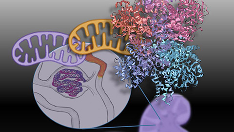
Mitochondria shape kidney cell function
Researchers at the University of Washington, Seattle present the first quantitative comparison of mitochondrial interactomes between two epithelial cell types in the kidney.
Long-chain polyunsaturated fatty acids linked to postoperative delirium risk
Researchers show that altered lipid metabolism may contribute to postoperative delirium, a condition linked to increased risk for long-term cognitive decline. The study explores potential disease mechanisms, which have yet to be understood.
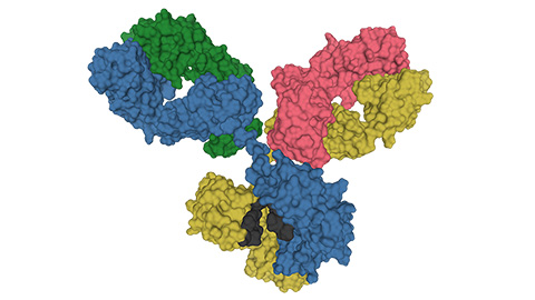
Glycosylation patterns across antibody isotypes distinguish tuberculosis states
Researchers at Taipei Medical University present the first site-specific glycosylation analysis of immunoglobulins in elderly tuberculosis patients.

Blood glycome possibly predicts lifespan
Researchers at the University of Santiago de Compostela show that total serum N-glycome can predict mortality independent of traditional risk factors.
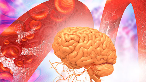
Building a better model for drug delivery across the blood–brain barrier
Industry and academic scientists collaborated to develop a rat with humanized iron-transport receptors, enabling research into iron homeostasis and drugs that cross the brain’s barrier.