Finding a missing piece for neurodegenerative disease research
Research led by the University of Michigan has provided compelling evidence that could solve a fundamental mystery in the makeup of fibrils that play a role in Alzheimer’s, Parkinson’s and other neurodegenerative diseases.
“We’ve seen that patients have these fibril structures in their brains for a long time now,” said Ursula Jakob, senior author of the new study. “But the questions are what do these fibrils do? What is their role in disease? And, most importantly, can we do something to get rid of them if they are responsible for these devastating diseases?”

Although the new finding does not explicitly answer those questions, it may provide a missing piece of the puzzle for researchers that are trying to understand how these diseases work at a molecular level. And it’s clear that this more intimate understanding is needed, given the lack of Alzheimer’s treatment options, Jakob said.
The Food and Drug Administration has approved three new drugs for Alzheimer’s disease since 2021, but that was preceded by a 17-year stretch without any new approvals despite hundreds of clinical trials (even now, there are more than 100 drug candidates being evaluated).
“Given all these unsuccessful clinical trials, we must still be missing some important pieces of this puzzle,” said Jakob, a professor in the U-M Department of Molecular, Cellular, and Developmental Biology. “So the fundamental research that we and many others around the world are doing is critically necessary if we ever want to treat, much less eliminate, these terrible diseases.”
The mystery density
Researchers have long known fibrils—tiny tendrils assembled from invisibly small building blocks called amyloid proteins—are connected to a number of neurodegenerative diseases. But important questions have lingered about how these structures build up in the body and how they affect the progression of these disorders.
Our understanding of the fibrils continues to develop as scientists introduce new tools and methods to probe the structures more intimately. One of those innovations is known as cryogenic electron microscopy, or cryo-EM.
“This is a very sophisticated technique,” Jakob said. “With it you can see what these fibrils look like in great detail.”
In 2020, an international team led by researchers in Cambridge using cryo-EM discovered a mysterious mass inside fibrils that were recovered from patients with a neurodegenerative disease called multiple system atrophy.
Although researchers could characterize the fibrils down to the individual amino acid units that build up the larger protein structure, there remained an unknown material running along the length of fibrils.
“It was right in the middle of the fibril and they had no idea what it was,” Jakob said. “They called it a ‘mystery density.'”
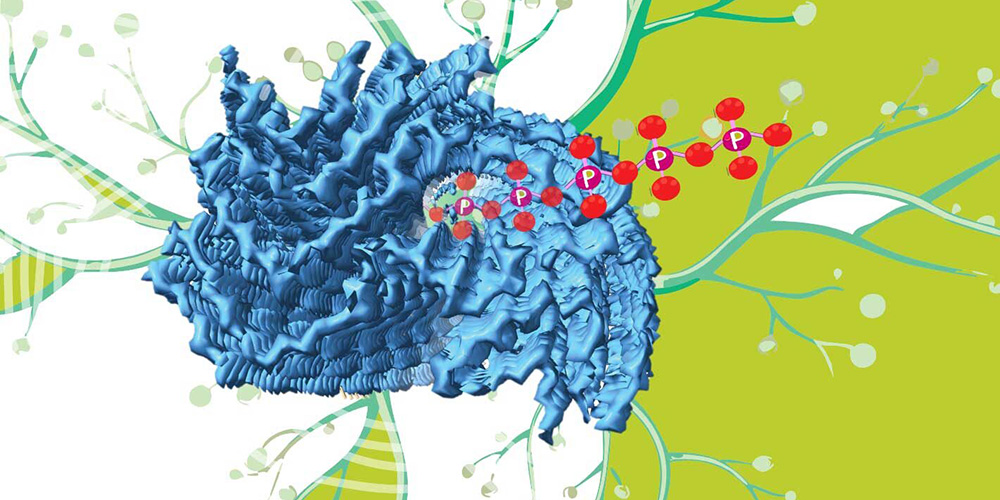
Now, Jakob and her colleagues have shown that a ubiquitous biological polymer called polyphosphate could be that mystery density.
The team reported its findings in the journal PLOS Biology.

New science, ancient molecule
Polyphosphate is a molecule found in every living thing today and has been used by organisms throughout the eons of evolution, Jakob said. It is also thought to have links to several neurodegenerative conditions thanks to laboratory experiments performed by Jakob and other scientists.
For example, her team showed that polyphosphate helps stabilize fibrils and reduces their destructive potential against lab cultured neurons. Other researchers have shown that the amount of polyphosphate in rat brains decreases with age.
These results imply polyphosphate could be important in protecting humans against neurodegenerative diseases. Still, scientists lacked direct evidence that it was.
“You can do a lot of things in test tubes,” Jakob said. “The question is which are genuinely relevant in the human body.”
The human brain, however, is an incredibly complex environment. Scientists have yet to design an experiment that can clearly elucidate polyposphate’s role in it.
But scientists did have precise, 3D structures of real fibrils from humans thanks to earlier research. By creating computer models of those structures, Jakob and her team could run simulations that asked how polyphosphate would interact with a fibril. They found that it fit the mystery density very well.
They then took it a step further and tweaked the structure of the fibril, changing the amino acids that bordered the mystery density. When they tested these fibril variants, they found that polyphosphate was no longer associated with them and no longer protected neurons against the fibrils’ toxicity.
“Because we’re unable to extract polyphosphate from patient-derived fibrils—it’s just not technically possible—we can’t say for sure that it is really the mystery density,” Jakob said. “What we can say is that we have very good evidence that the mystery density fits polyphosphate.”
Their work leads to the hypothesis that finding a way to maintain proper polyphosphate levels in the brain could possibly slow the progress of neurodegenerative disease. But proving that will still take large investments of time and money, Jakob said, and there will likely be new mysteries to be solved along the way.
“I would say we are still at a very early stage. It’s only very recently that it became clear that there are additional components in these fibrils,” she said. “These components may play a huge role or they might not play any role at all. But only if we have the pieces of the puzzle in place, can we hope to be able to successfully fight these hugely devastating diseases.”
This article is republished from the Michigan News. Read the original here.
Enjoy reading ASBMB Today?
Become a member to receive the print edition four times a year and the digital edition monthly.
Learn moreGet the latest from ASBMB Today
Enter your email address, and we’ll send you a weekly email with recent articles, interviews and more.
Latest in Science
Science highlights or most popular articles

Life in four dimensions: When biology outpaces the brain
Nobel laureate Eric Betzig will discuss his research on information transfer in biology from proteins to organisms at the 2026 ASBMB Annual Meeting.

Fasting, fat and the molecular switches that keep us alive
Nutritional biochemist and JLR AE Sander Kersten has spent decades uncovering how the body adapts to fasting. His discoveries on lipid metabolism and gene regulation reveal how our ancient survival mechanisms may hold keys to modern metabolic health.

Redefining excellence to drive equity and innovation
Donita Brady will receive the ASBMB Ruth Kirschstein Award for Maximizing Access in Science at the ASBMB Annual Meeting, March 7–10, just outside of Washington, D.C.

Mining microbes for rare earth solutions
Joseph Cotruvo, Jr., will receive the ASBMB Mildred Cohn Young Investigator Award at the ASBMB Annual Meeting, March 7–10, just outside of Washington, D.C.

Fueling healthier aging, connecting metabolism stress and time
Biochemist Melanie McReynolds investigates how metabolism and stress shape the aging process. Her research on NAD+, a molecule central to cellular energy, reveals how maintaining its balance could promote healthier, longer lives.

Mapping proteins, one side chain at a time
Roland Dunbrack Jr. will receive the ASBMB DeLano Award for Computational Biosciences at the ASBMB Annual Meeting, March 7–10, just outside of Washington, D.C.

