MCP: How radiotherapies
vanquish cancer cells

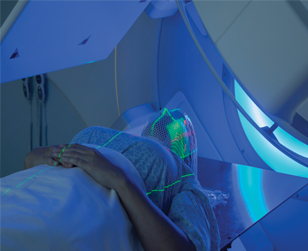
Tumor-shredding therapies aren't created equal. When someone with cancer decides to treat the cellular aberration with radiation therapy, as more than 50 percent of cancer patients now do, their primary options are X-rays or particle beams. Despite being available at only a handful of treatment centers worldwide, particle beams, which use protons or carbon ions, possess a number of benefits over traditional X-rays.
The effects of the beams on the mechanisms that govern signaling within cells remain largely unexplored. A recent paper in the journal Molecular & Cellular Proteomics sheds some light on this matter.
To better understand the regulatory effects that the particle-based techniques have on the structure and signaling pathways of cancerous cells, researchers at the German Cancer Research Center, Heidelberg University and Heidelberg Ion Beam Therapy Center applied a combination of high-resolution mass spectrometry and SILAC to irradiated human lung adenocarcinoma cells. SILAC is short for stable isotope labeling by amino acids in cell culture; by incorporating amino acids labeled with heavy isotopes into cell cultures and comparing those cells’ mass spectrometry peaks to those of identical, untreated cell cultures, researchers can examine the effects of an outside agent, such as radiation, on the protein makeup of a culture.
“The research interest is just finding out what radiation is doing to cells, to humans and to tissues,” says senior author Martina Schnölzer at the center’s Functional Proteome Analysis unit. “As a chemist, it was really interesting to work together with the radiation oncologists, because my background is more proteomics and not medicine.”
Whereas X-rays destroy cancer cells by inflicting strand breaks on DNA’s double-helix structure, which can be fixed by DNA repair mechanisms, protons and carbon ions tear the genetic structure apart with complex double-strand breaks, which cannot be repaired. This gives proton and carbon ion beams a larger relative biological effectiveness than X-rays, meaning they kill more cancer cells than X-rays do at the same dose. Additionally, particle beams deposit their energy in a more focused manner than X-rays, damaging to a lesser extent the healthy tissues surrounding tumors.
The researchers separated cultures of the human lung cancer cell line into two groups that were fed amino acids with heavy isotopes or light isotopes and irradiated subgroups of the heavy-labeled cells with x-rays, carbon ions or protons. They then performed a phosphopeptide enrichment to increase the concentration of phosphorylated proteins in each sample before subjecting the cells to mass spectrometry to identify changes that had occurred at the protein structure level and which sites had phosphate molecules added or removed. The addition or removal of these molecules is a key indicator that cells are attempting to mitigate damage and is known in aggregate as the phosphoproteome.
While the researchers observed only limited effects on lung cells at the protein level, they noticed altered phosphorylation regulation on 181 different protein sites, or residues, 151 of which had not been previously been known to be affected by irradiation.
“We’ve looked at differential quantification of the proteins and found that there is little happening as an initial event as a consequence of radiation, whereas the phosphoproteome is massively (dysregulated),” says senior co-author Amir Abdollahi at the center’s Division for Molecular and Translational Radiation Oncology. This information will be helpful for designing future studies that examine the effects of radiotherapy, ionizing radiation and space radiation on cellular signaling processes, he says.
Future work for Schnölzer and Abdollahi will involve looking at the effects of radiation on a longer timescale to understand late effects of irradiation and examining its effects on pathways other than phosphorylation signaling.
Enjoy reading ASBMB Today?
Become a member to receive the print edition four times a year and the digital edition monthly.
Learn moreGet the latest from ASBMB Today
Enter your email address, and we’ll send you a weekly email with recent articles, interviews and more.
Latest in Science
Science highlights or most popular articles
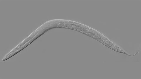
How smelling death alters worm behavior
Researchers have found that the roundworm C. elegans can smell death, and it changes how the worms behave, reproduce and age.
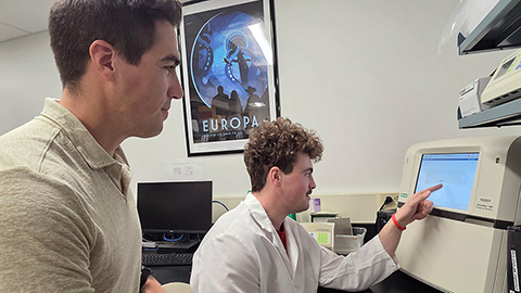
A chance encounter with the lab
Payton Stevens never planned to become a pancreatic cancer researcher. A temporary job set him on a path from rural Kentucky to leading research on Wnt signaling and metastasis, where he now pairs discovery with mentorship and science advocacy.
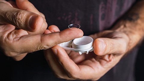
Light-activated small molecule could transform eye infection treatment
Contact lenses raise the risk of infectious keratitis, a leading cause of blindness worldwide. A biotech company is commercializing a light-activated therapy using a ROS-generating molecule to rapidly kill microbes in the cornea to preserve vision.

The molecular orchestra of memory
Calcium, calmodulin and calcium/calmodulin-dependent kinase II form a molecular axis that turns fleeting neural activity into lasting memories. New research shows how memories are stabilized, and possibly even protected or repaired.
Differences in pili structure modulate bacterial behavior
Researchers demonstrate how small changes in the structure of hair-like protein appendages can affect the behavior of Acinetobacter bacteria.

Cholesterol regulatory genes predict liver transplant outcomes
Researchers identify a link between cholesterol-regulating genes and liver transplant success, which could improve donor screening and patient outcomes.