Cow born in Japan after removal, replacement of placental cells
Researchers at Hokkaido University have found that cow embryos from which placenta-forming cells had been removed can regrow those cells, form a placenta and successfully gestate. The scientists recently published their results, which provide insight into the regenerative capacity of mammalian embryos, in the Journal of Biological Chemistry.
All mammalian embryos follow the same blueprint in the first week of development: After being fertilized, a zygote divides into two cells, which quickly become four, eight, then 16 cells that specialize into an inner cell mass and outer cells that are known individually as trophoblasts and collectively as the trophectoderm.
Nanami Kohri, the lead author on the paper, was intrigued by the fact that mouse embryos in which the trophoblasts — which differentiate to form the placenta — had been removed were much less successful in regenerating a placenta than bovine embryos that also had trophoblasts removed.
“Although isolated inner cell masses in both mice and cattle underwent trophectoderm regeneration, they were significantly different in terms of regeneration efficiency, marker protein distribution and expression status of key genes,” he said. “Surprisingly, a calf was successfully delivered after the transfer of the reformed inner cell mass to the surrogate mother, but no descendants were obtained from reformed inner cell masses in mice.”

Kohri and his colleagues at the Laboratory of Animal Breeding and Reproduction previously had isolated bovine inner cell masses from embryos at the early blastocyst stage to find where the genes that give rise to the trophectoderm were being expressed. Other groups had shown that cells positioned at the outer margin of the inner cell mass could be transformed into trophectoderm in mouse embryos.
To understand why the bovine embryos had more success regenerating placental cells than the murine embryos, the researchers at Hokkaido University investigated the expression of the gene SOX17, which creates a protein that regulates cell specialization in development. They found that the expression of SOX17 varied significantly between the two species and was localized to the trophectoderm cells that had been originally absent in murine embryos, which might explain the weaker regenerative capacity.
Kohri and colleagues plan to investigate what drives the differences in embryonic protein expression among mammals as they continue to monitor their calf, which is now 23 months old and healthy.“It has been suggested that the molecular basis of determining cellular divisions and localization in development differs among species,” Kohri said. “In the future, we will have to use our experimental system to evaluate trophectoderm regeneration from the reformed inner cell masses in mice and cattle.”
Enjoy reading ASBMB Today?
Become a member to receive the print edition four times a year and the digital edition monthly.
Learn moreGet the latest from ASBMB Today
Enter your email address, and we’ll send you a weekly email with recent articles, interviews and more.
Latest in Science
Science highlights or most popular articles
Uncovering the molecular roots of fatty liver disease
Physician–scientist Silvia Sookoian discusses her path from hepatitis C care to MASLD research, her use of multi-omics to study steatotic liver disease, and how lipid metabolism and genetics are reshaping understanding of MASH and liver health.
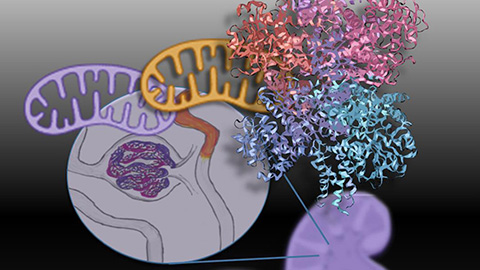
Mitochondria shape kidney cell function
Researchers at the University of Washington, Seattle present the first quantitative comparison of mitochondrial interactomes between two epithelial cell types in the kidney.
Long-chain polyunsaturated fatty acids linked to postoperative delirium risk
Researchers show that altered lipid metabolism may contribute to postoperative delirium, a condition linked to increased risk for long-term cognitive decline. The study explores potential disease mechanisms, which have yet to be understood.
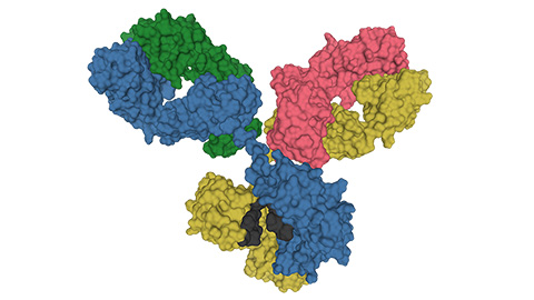
Glycosylation patterns across antibody isotypes distinguish tuberculosis states
Researchers at Taipei Medical University present the first site-specific glycosylation analysis of immunoglobulins in elderly tuberculosis patients.

Blood glycome possibly predicts lifespan
Researchers at the University of Santiago de Compostela show that total serum N-glycome can predict mortality independent of traditional risk factors.
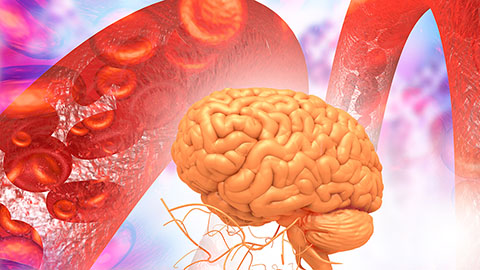
Building a better model for drug delivery across the blood–brain barrier
Industry and academic scientists collaborated to develop a rat with humanized iron-transport receptors, enabling research into iron homeostasis and drugs that cross the brain’s barrier.