How is myelin made?

Myelin is the protective lipid sheath wrapped around a nerve. It functions as an insulator, akin to the protective coating on a wire, speeding up electrical transmission of signals along a neuron. Myelin also plays a role in maintaining the health of neurons. Myelin function is dysregulated in many neurological disorders, including multiple sclerosis.
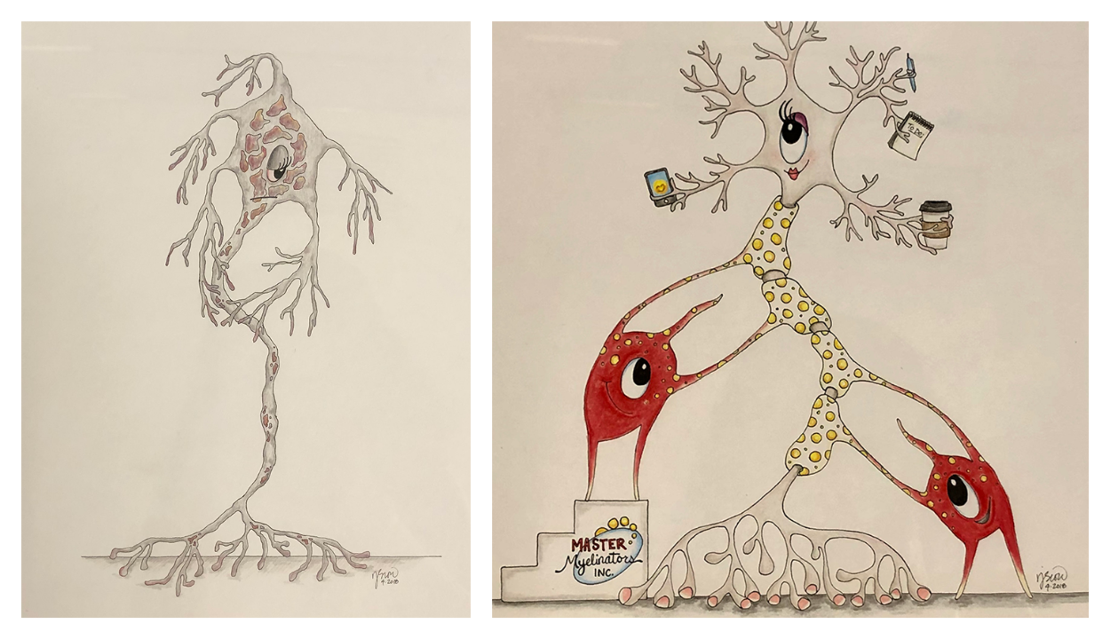
Oligodendrocytes are the myelin-producing cells of the central nervous system. The myelin sheath around a neuron is part of an oligodendrocyte’s plasma membrane, and a single oligodendrocyte can myelinate as many as 50 neurons. During myelination, an oligodendrocyte stretches out tubes of membrane in search of a neuron. When it finds one, it sends the necessary building materials down the tubes and, still operating from a distance, assembles a myelin sheet around the neuron: Composition, number of wraps and total coverage all matter. A myelinated neuron that loses its coating cannot transmit electrical signals properly, leading to loss of muscle control and other neurological problems.
The myelin sheath is mostly made of lipids, including sphingolipids, which are critical to myelin’s structure and function. The enzyme serine palymitoyltransferase, or SPT, produces the backbone of all sphingolipids, and the membrane-bound protein ORMDL monitors sphingolipid levels and regulates SPT activity. ORMDL’s activity must be precise: Too little sphingolipid production impedes myelination, and too much can be toxic.
Binks Wattenberg, a professor of biochemistry and molecular biology at Virginia Commonwealth University, studies membrane biogenesis and now focuses on lipid biogenesis. “I am very curious about how the cell knows when to make sphingolipid and when to stop,” Wattenberg said. “I think ORMDL might be the key to answering that question.”
Wattenberg’s next-door lab neighbor, Carmen Sato–Bigbee, a professor in the same department, studies myelination, with a focus on oligodendrocytes. The two joined forces to study the role of sphingolipid biosynthesis in myelination in developing brains, and reported their results in the Journal of Lipid Research.
To uncover the dynamics of sphingolipid content and synthesis during myelination, Wattenberg and Sato–Bigbee’s team worked with newborn rat brains, because peak myelination occurs directly after birth. Only one in five cells in the brain is an oligodendrocyte, so the team isolated these myelin-producing cells for their experiments.
The researchers found that a large portion of the sphingolipids present in oligodendrocytes during myelination have an atypically long backbone — an 18-carbon chain instead of a 16-carbon chain. “The 18-carbon chain backbone points to a change in lipid composition during myelination, which might explain the insulating properties of myelin,” Wattenberg said. “In future work, we want to look at the role of each type of sphingolipid in myelination.”
The study also found that SPT activity increases for the first few days of myelination and then begins to decrease. ORMDL activity is not measurable, but the team deduced that ORMDL isoform expression varies over time. These findings pave the way for future experiments.
“The control of sphingolipid biosynthesis is key to myelination, and understanding how this process works will enable us to alter it in future treatments,” Wattenberg said. “Our pie-in-the-sky goal is to understand sphingolipid biosynthesis so well that we can reprogram oligodendrocytes and reverse demyelination in degenerative myelination diseases like MS.”
Enjoy reading ASBMB Today?
Become a member to receive the print edition four times a year and the digital edition monthly.
Learn moreGet the latest from ASBMB Today
Enter your email address, and we’ll send you a weekly email with recent articles, interviews and more.
Latest in Science
Science highlights or most popular articles
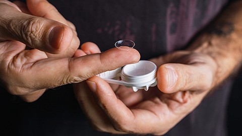
Light-activated small molecule could transform eye infection treatment
Contact lenses raise the risk of infectious keratitis, a leading cause of blindness worldwide. A biotech company is commercializing a light-activated therapy using a ROS-generating molecule to rapidly kill microbes in the cornea to preserve vision.

The molecular orchestra of memory
Calcium, calmodulin and calcium/calmodulin-dependent kinase II form a molecular axis that turns fleeting neural activity into lasting memories. New research shows how memories are stabilized, and possibly even protected or repaired.
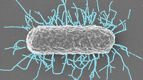
Differences in pili structure modulate bacterial behavior
Researchers demonstrate how small changes in the structure of hair-like protein appendages can affect the behavior of Acinetobacter bacteria.

Cholesterol regulatory genes predict liver transplant outcomes
Researchers identify a link between cholesterol-regulating genes and liver transplant success, which could improve donor screening and patient outcomes.

Lipid signatures for a rare neurological disorder
Researchers find distinct lipid patterns linked to a rare autoimmune neurological disorder, offering hope for effective targeted therapies for patients.
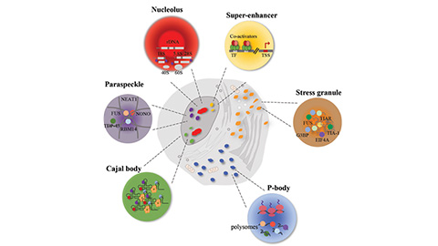
Disease-linked mutations disrupt protein phase behavior
Researchers find that pathogenic missense mutations are enriched threefold in phrase-separating intrinsically disordered regions of proteins.