Breaking dogma?
When a molecular entity comes along that appears to affect cancer biology, immunology and neurobiology – as well as turn conventional molecular biology wisdom on its head – people tend to sit up and notice. They start thinking up ways to exploit the molecular entity for diagnostic and therapeutic purposes. They may even put forward $17 million to investigate this molecular entity, as did officials at the National Institutes of Health’s Common Fund program in 2013.
 If research confirms RNA molecules are transferred via vesicles between cells, the phenomenon will represent a paradigm shift in molecular biology.
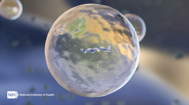
That’s what is happening with extracellular vesicles that carry RNA. RNA long has been thought to be a molecule imprisoned within a cell. Proteins, like hormones, always have been thought to be the ones to do long-distance communications. If more research confirms that RNA is transferred from one cell to another through extracellular vesicles and causes the recipient cell to change its behavior, this phenomenon will represent a paradigm shift in biology.
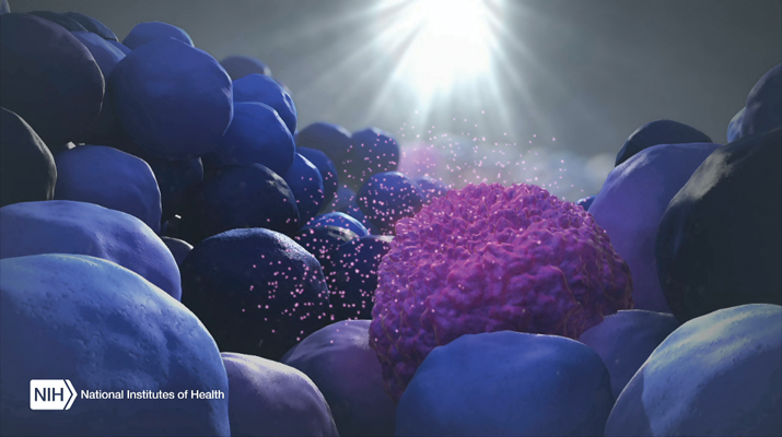
Researchers have documented RNA-containing extracellular vesicles triggered by environmental stimuli coming off brain tumor cells, inflammatory cells and neurons. They don’t contain a random jumble of RNA: Specific types of RNA are found in the vesicles. They tend to be small, noncoding RNAs as well as microRNAs. The assortment of RNAs hints that the process of making these vesicles is highly regulated.
The vesicles can be found in every body fluid, such as blood, urine and semen, so researchers easily can envision using extracellular vesicles as diagnostic and therapeutic agents. Collecting the body fluids and using the vesicles in them to diagnose diseases and illnesses is a tantalizing idea, because it’s easier to do than more invasive procedures.
“It’s a very exciting field right now,” says Johan Skog at Exosome Diagnostics. “But we have to be careful to not get too carried away.” Michiel Pegtel at VU University in The Netherlands concurs. “You want to think big when talking about these small vesicles,” he says. “But you want to be very cautious.”
The skepticism is warranted, because “the functionality of these RNAs in recipient cells is a question for debate,” acknowledges Xandra Breakefield at the Massachusetts General Hospital. “Right now, there is very little conclusive data in that area. It’s just the idea that wouldn’t it be amazing if RNA could actually communicate?”
“Anything is possible now”
If RNA does manipulate far-flung cells through extracellular vesicles, the implications for cell biology are profound. “The dogma was (that) we have genetic information in multicellular organisms, like ourselves, as DNA. It’s inside the nucleus, and it’s nicely protected there,” says Pegtel. Any movement of genetic information happened within the confines of the cells in multicellular organisms using RNA.
“Bacteria might do all kinds of weird things to exchange genetic information with each other, but we, higher organisms, didn’t do that,” says Pegtel. “That was the dogma.” Now, with the discovery of these extracellular vesicles containing RNA, Pegtel adds, “it’s a paradigm shift in how we think multicellular organisms function. That’s where the excitement comes from.”
One exciting idea is that microbial and plant-based foods come into our bodies with their own baggage of extracellular vesicles. The issue of cross-species interactions is “an extremely interesting and important question to ask,” says Pegtel. “For instance, do we communicate with our commensal bacteria in the gut, and does our genome have an effect on the genomes of bacteria? This is one of the reasons why there is so much skepticism – because, potentially, anything is possible now.”
Making them work
Besides remodeling our understanding of cell-cell communications, researchers think extracellular vesicles can do some work for them. For example, these extracellular vesicles could be used as biomarkers. Louise Laurent at the University of California, San Diego, says genetic testing on fetal DNA found in maternal blood opens up the possibility to look for fetal RNA in extracellular vesicles floating in maternal blood. “RNA is much more dynamic than DNA,” she says. With DNA, “we usually use it to check if there’s a genetic abnormality, but RNA, we reason, could change with different complications of pregnancy. They might even change prior to the onset of signs or symptoms that we could clinically identify.”
Relying on extracellular-vesicle cargo as diagnostic biomarkers will be a reality shortly. Exosome Diagnostics, where Skog works, is gearing up to release a prostate cancer test that detects RNA in vesicles found in urine. The timeline for having the diagnostic test on the market is “months,” says Skog.
Researchers also are looking to exploit these vesicles for therapeutic purposes. Therapeutics based on vesicles with RNA is a very appealing idea, because the vesicles might be made to target specific cells carrying RNA with a particular set of instructions. Using nanoscale lipid vesicles as carriers for drugs is not a new idea, but the discovery of naturally occurring vesicles that target certain cell types gives researchers a new form of vesicles that come endowed with the bells and whistles needed for targeting and delivery of cargo. Richard Kraig at The University of Chicago Medical Center rattles off the attributes of the vesicles: They are nontoxic, conserved among species, targeted for specific cells and carry potent signaling molecules.
However, therapeutic applications of these vesicles are a longer way off, say experts. “Five years away from therapeutics – that’s my dream,” says Kraig. For his work, Kraig is using naturally occurring exosomes to encourage myelination in animal models of neurodegenerative disorders, such as multiple sclerosis.

What are we talking about?
To understand why caution must temper the enthusiasm over extracellular vesicles, simply consider the fact that researchers in the field are still trying to agree on a name for what they are studying. “The terminology has not been sorted out yet,” says Breakefield.
The problem is that the field is young and still riddled with fundamental questions. It was only in 2007 that researchers were able to demonstrate that extracellular vesicles contained RNA. So the questions began: Why do cells bundle RNA into lipid vesicles? Are all vesicles created the same? How are they targeted to receiving cells?
Extracellular vesicles are among a cadre of other vesicles found in the body, so it’s critical to be clear exactly which kind of vesicle is being discussed. Right now, Skog says, the vesicles “have different names depending on what the (research) group is working on. Everyone thinks they are their favorite vesicles, but it’s really a mixed bag of vesicles from different origins, and there’s no way now of knowing where they are coming from after they have left the cell.”
Several different names, such as “exosomes” or “microvesicles,” have been used in the literature to describe the lipid-encapsulated particles. These days, most researchers in the field of vesicles call the RNA-containing vesicles “exosomes.” But the waters remain murky, because different people give different rationales for calling their vesicles exosomes.
One popular naming procedure relies on the molecular mechanism: Label vesicles coming from the endosomes as exosomes and those vesicles blebbing off the plasma membrane as microvesicles. But this definition runs into problems, because the mechanisms by which these vesicles are made still remain to be elucidated unequivocally. Fans of the mechanistic form for naming vesicles say that the exosomes and microvesicles can be distinguished from one another by different protein markers on the surfaces. Exosomes are the vesicles coming from the multivesicular pathways that bear tetraspanin proteins like CD9, CD81 and CD63. Microvesicles supposedly don’t have these markers, because they bleb from the cell surface.
“We know that’s not true,” counters Douglas Taylor at Exosome Sciences. “Almost any vesicle that comes from a tumor cell has a tetraspanin, whether it’s blebbed from the surface or if it comes from the endocytic pathway. So that’s not really a good definition.”
Furthermore, “there is no data that demonstrates that the mechanism of vesicle budding at the plasma membrane is mechanistically different from that which occurs in the endosome,” adds Stephen Gould at Johns Hopkins University. “They might be simply two ends of the same spectrum.”
Some researchers want to use size as the defining factor. Exosomes are vesicles that are smaller than 200 nanometers, and microvesicles are lipid bodies that are 500 nanometers and bigger, they say. But the size has no bearing on the biological effect elicited by the vesicles.
Until there’s a better understanding of the biology, some researchers, like Breakefield and Gould, suggest that the best course of action for now is to use the more generic term – extracellular vesicles. “In lieu of having good, strong experimental data and detailed mechanistic hypotheses tested rigorously by multiple labs, the models are mostly imaginations of what we think is probably going on rather than empirically derived, well-accepted scientific facts,” says Gould, who is the president of the American Society for Exosomes and Microvesicles.
What are you looking at?
Besides coming up with a name, researchers also have to figure out how to ensure they are looking at the same entities. “There are a large number of papers where people demonstrate some biological functions of exosomes. But it’s very hard to compare them, because there are no standards for workflows,” says Alexander “Sasha” Vlassov at Thermo Fisher Scientific, whose group is involved in developing tools to study extracellular vesicles. “It’s still the wild West.”
Indeed, trying to compare results is a problem. “One of the things impeding the advancement in this field is that not only there isn’t well-defined terminology or nomenclature, but there’s also no well-established protocols that people can agree on” for isolating, purifying and analyzing extracellular vesicles, says Danilo Tagle of the National Center for Advancing Translational Sciences at the National Institutes of Health.
The conventional tools for isolating extracellular vesicles are ultracentrifugation and density gradient separations. But companies have jumped in with kits. “There are a zillion different types of kits that try to isolate vesicles from different cell supernatants with different types of antibodies or even with polyethylene glycol,” says Jan Lötvall at Gothenburg University in Sweden, who is the president of the International Society of Extracellular Vesicles.
Tagle says one way in which the NIH Common Fund program in extracellular RNA is hoping to help the field move forward is to “come out with a best set of recommendations” for analyzing extracellular vesicles.
Lots of questions
A looming issue is that no one is sure yet how to demonstrate the scale of importance of extracellular vesicles. “We still don’t really know how to block the secretion of exosomes without disrupting the whole cell. It might not even be possible,” says Pegtel.
He gives the example of mitochondria. “You can’t prove the importance of mitochondria by saying, ‘I’m going to knock out mitochondria in cells and then show that they are important.’ It’s a little bit of a catch-22,” he notes.
Lötvall’s group has seen “the translation of mouse proteins in the recipient human cells. It didn’t change the phenotype of the recipient cell, but it showed that the RNA was actually transferred in an intact form and was functional in the recipient cell. It wasn’t destroyed during the uptake process.” But Lötvall says he hasn’t seen evidence that proves that a mixture of RNAs in an extracellular vesicle elicits a change in the recipient cells within the same organism. He says, “The killer experiment, to prove or disprove the importance of extracellular RNA in normal physiology, still remains to be done.”
Experts keep returning to the fact that they have more questions than answers at this point. Where are the vesicles made inside the cell? How do certain RNAs get packaged into them? How often are the vesicles made? What aspect of the pathway goes wrong in different diseases? “There is an insane amount of questions at the cell biological level that we still need to answer,” says Pegtel.
And the biggest question of all is whether the buzz surrounding extracellular vesicles will hold up when one question in particular gets scrutiny. As Pegtel put it: “How big is the role of exosomes, and how useful are they?”
Enjoy reading ASBMB Today?
Become a member to receive the print edition four times a year and the digital edition monthly.
Learn moreGet the latest from ASBMB Today
Enter your email address, and we’ll send you a weekly email with recent articles, interviews and more.
Latest in Science
Science highlights or most popular articles

Mapping proteins, one side chain at a time
Roland Dunbrack Jr. will receive the ASBMB DeLano Award for Computational Biosciences at the ASBMB Annual Meeting, March 7–10, just outside of Washington, D.C.

Exploring the link between lipids and longevity
Meng Wang will present her work on metabolism and aging at the ASBMB Annual Meeting, March 7-10, just outside of Washington, D.C.

Defining a ‘crucial gatekeeper’ of lipid metabolism
George Carman receives the Herbert Tabor Research Award at the ASBMB Annual Meeting, March 7–10, just outside of Washington, D.C.

The science of staying strong
Muscles power every movement, but they also tell the story of aging itself. Scientists are uncovering how strength fades, why some species resist it and what lifestyle and molecular clues could help preserve muscle health for life.

Bacteriophage protein could make queso fresco safer
Researchers characterized the structure and function of PlyP100, a bacteriophage protein that shows promise as a food-safe antimicrobial for preventing Listeria monocytogenes growth in fresh cheeses.

Building the blueprint to block HIV
Wesley Sundquist will present his work on the HIV capsid and revolutionary drug, Lenacapavir, at the ASBMB Annual Meeting, March 7–10, in Maryland.

