Nanoplastics may help set the stage for Parkinson’s risk

Parkinson’s disease and related dementias have been on the rise worldwide. These disorders are marked by an abnormal buildup of the protein alpha-synuclein in the brain. The factors leading to this buildup of alpha-synuclein are unknown. Research points to a potential role for environmental factors.
Small bits of plastic are widely found throughout the environment, including food and water supplies. Microplastics are plastic particles smaller than 5 mm in diameter—tinier than a sesame seed; nanoplastics are less than 1 μm, too small to be seen by the human eye. At least one previous study found that particles of polystyrene and other plastics can be detected in the blood of most healthy adults. Single-use polystyrene products—like plastic cups, utensils, and foam packing—are widespread environmental waste. But despite their ubiquity, the potential health consequences of these plastics are only beginning to be studied and understood.

Previous studies found evidence that alpha-synuclein’s activities can be affected by polystyrene and other particles. An international research team led by Dr. Andrew B. West of Duke University decided to take a closer look at the effects that nanoplastics might have on nerve cells and the brain. The scientists explored interactions between alpha-synuclein and polystyrene nanoplastics both in lab dishes and in mice. Results were reported on November 17, 2023, in Science Advances.
The researchers first showed that human alpha-synuclein binds readily to polystyrene nanoplastics in a test tube. This binding led to the formation of abnormal alpha-synuclein structures called fibrils, a hallmark of Parkinson’s disease and related dementias.
The scientists next examined how alpha-synuclein fibrils and nanoplastics behave with cultured brain cells, or neurons. They found that both the fibrils and the plastics can enter neurons via endocytosis, in which the cell’s outer membrane engulfs targeted items. Once inside, both the fibrils and the plastics entered the cell’s lysosomes, membrane-bound organelles that serve as cellular garbage disposals. The researchers found that nanoplastics disrupted lysosome activities, slowing the breakdown of harmful clumps of alpha-synuclein.
The team next looked at how polystyrene nanoplastics and alpha-synuclein interact in the mouse brain. They found that the nanoplastics and alpha-synuclein fibrils also interacted there, which increased the spread of abnormalities across interconnected brain regions. Neurons in the brain’s substantia nigra region were especially affected. This brain region helps to control movement and is damaged in Parkinson’s disease and related dementias.
Taken together, these findings point to previously unrecognized interactions that could contribute to Parkinson’s disease risk and progression. Further research is needed to study how these interactions affect disease development and whether other types of plastics have similar effects.
“Numerous lines of data suggest environmental factors might play a prominent role in Parkinson’s disease, but such factors have for the most part not been identified,” West explains. “Our study suggests that the emergence of micro and nanoplastics in the environment might represent a new toxin challenge with respect to Parkinson’s disease risk and progression.”
This story originally appeared on the NIH Research Matters website. Read the original article.
Enjoy reading ASBMB Today?
Become a member to receive the print edition four times a year and the digital edition monthly.
Learn moreGet the latest from ASBMB Today
Enter your email address, and we’ll send you a weekly email with recent articles, interviews and more.
Latest in Science
Science highlights or most popular articles

Flipping lipids and slime molds
A dull first job nearly pushed JBC associate editor Todd Graham out of science. Then a slime mold project changed his path. Now, he studies membrane biology and reflects on discovery, persistence and mentoring through uncertainty.
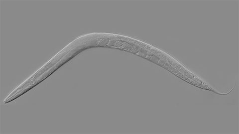
How smelling death alters worm behavior
Researchers have found that the roundworm C. elegans can smell death, and it changes how the worms behave, reproduce and age.
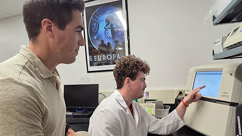
A chance encounter with the lab
Payton Stevens never planned to become a pancreatic cancer researcher. A temporary job set him on a path from rural Kentucky to leading research on Wnt signaling and metastasis, where he now pairs discovery with mentorship and science advocacy.
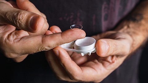
Light-activated small molecule could transform eye infection treatment
Contact lenses raise the risk of infectious keratitis, a leading cause of blindness worldwide. A biotech company is commercializing a light-activated therapy using a ROS-generating molecule to rapidly kill microbes in the cornea to preserve vision.

The molecular orchestra of memory
Calcium, calmodulin and calcium/calmodulin-dependent kinase II form a molecular axis that turns fleeting neural activity into lasting memories. New research shows how memories are stabilized, and possibly even protected or repaired.
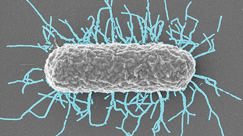
Differences in pili structure modulate bacterial behavior
Researchers demonstrate how small changes in the structure of hair-like protein appendages can affect the behavior of Acinetobacter bacteria.