Patients lose weight in trials of schizophrenia drug

There remains a significant need for new therapeutic options for the treatment of patients with, often debilitating, psychiatric disorders. Current therapies are frequently associated with side effects, including unexplained weight gain in about one-third of patients. Breakthroughs in brain research and the discovery of potential novel drug targets are leading to the development of a new generation of candidate therapies that are providing early signs of hope for these patients.

Hence, when Michal Tomaszewski, an associate principal scientist at Merck & Co., and his team learned from their colleagues that patients treated with an investigational antipsychotic medicine lost weight in a Phase 2a clinical trial, they knew they needed to better understand the mechanism of weight decline.
In their recent paper published in the Journal of Lipid Research, the researchers record the results of their investigation, which showed the antipsychotic drug, a phosphodiesterase-10A, or PDE10A, inhibitor, significantly reduced the amount of stored fat in both white and brown adipose tissue in mice.
“Studying this transition lets us better understand the effect of the PDE10A inhibitor treatment on metabolism,” Tomaszewski said.
When white adipose tissue exhibits increased vasculature and metabolism, it is an indicator of its conversion into brown adipose tissue, or BAT. BAT cells burn up fat as heat energy, and increasing levels of these cells are often accompanied by weight loss. The researchers observed an increase in blood vessels and blood flow in white adipose tissue compared to a control.
“Specifically, the white adipose tissue showed enhanced blood vessel growth and increased consumption of fat for energy,” Tomaszewski said. “These changes were consistent with a transition to brown adipose tissue, which is known to be important in how we store and use energy, and therefore how we gain and lose weight.”
To see these effects, the researchers used magnetic resonance imaging in a novel way that helped them characterize the physiological changes in adipose tissue in response to the PDE10A inhibitor. They observed the transition of white adipose tissue into brown adipose tissue, which has higher energy metabolism. This explained the weight loss seen in PDE10A inhibitor-treated mice.
“This magnetic resonance imaging application provided detailed insights into metabolism and physiology not available using most other methods,” Tomaszewski said. “This technique may, in the future, be applied more broadly to evaluate the response of adipose tissue to additional experimental drug candidates.”
Tomaszewski believes this work resulted in multiple benefits. “Although imaging of mice in vivo was the focus of the study and made it possible to visualize the changing nature of the adipose tissue,” he said, “we were also able to perform comprehensive validation using genetic and protein analysis, which all aligned to reinforce our conclusions.”
Tomaszewski said he is optimistic that the magnetic resonance imaging methods used in this work “can be scaled to evaluate similar metabolic changes in people.”
Enjoy reading ASBMB Today?
Become a member to receive the print edition four times a year and the digital edition monthly.
Learn moreGet the latest from ASBMB Today
Enter your email address, and we’ll send you a weekly email with recent articles, interviews and more.
Latest in Science
Science highlights or most popular articles
Long-chain polyunsaturated fatty acids linked to postoperative delirium risk
Researchers show that altered lipid metabolism may contribute to postoperative delirium, a condition linked to increased risk for long-term cognitive decline. The study explores potential disease mechanisms, which have yet to be understood.
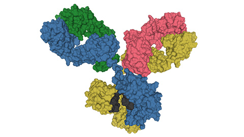
Glycosylation patterns across antibody isotypes distinguish tuberculosis states
Researchers at Taipei Medical University present the first site-specific glycosylation analysis of immunoglobulins in elderly tuberculosis patients.

Blood glycome possibly predicts lifespan
Researchers at the University of Santiago de Compostela show that total serum N-glycome can predict mortality independent of traditional risk factors.
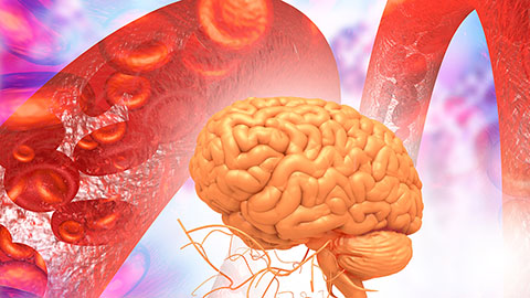
Building a better model for drug delivery across the blood–brain barrier
Industry and academic scientists collaborated to develop a rat with humanized iron-transport receptors, enabling research into iron homeostasis and drugs that cross the brain’s barrier.
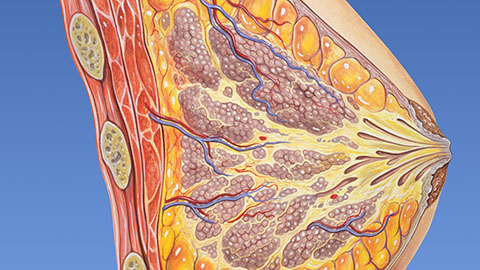
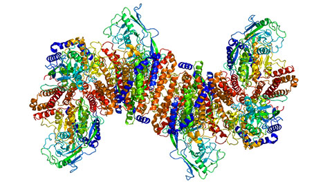
Fat synthesis enzyme crucial for milk fat and newborn growth
Researchers found that a deficiency of the fatty acid synthesis enzyme stearoyl-CoA desaturase-1 reduced mammary gland function during lactation and caused low birth weight in newborns that were fed milk from enzyme-deficient glands.
Flipping lipids and slime molds
A dull first job nearly pushed JBC associate editor Todd Graham out of science. Then a slime mold project changed his path. Now, he studies membrane biology and reflects on discovery, persistence and mentoring through uncertainty.